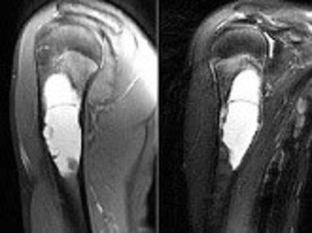
Unicameral bone cyst
A pseudo-tumor resembling a tumor, the unicameral bone cyst typically emerges at a young age, more commonly in males, often found on the femur but can affect any bone.
Cause
The exact cause of the unicameral bone cyst remains unknown, with suspicion pointing towards a local circulation disorder.
Symptoms
While asymptomatic in small sizes and discovered incidentally in adulthood, larger cysts may cause local pain due to microfractures. Complete fractures (pathological fractures) can occur from minimal trauma, attributed to thinning of the bone cortex. Active cysts near the growth plate may disrupt bone development, causing growth reduction or axial deviation
Diagnosis
Clinical history and examination may raise suspicion, confirmed by radiographic examination and complemented by MRI to observe possible cystic contents. Biopsy with microscopic examination is essential for confirmation.

Treatment
The approach to unicameral bone cysts varies based on size. Conservative methods involve infiltrating the cyst with slowly absorbed cortisone under guidance or surgically emptying the cavity, subsequently filling it with autologous or synthetic bone grafts. In cases of voluminous or load-bearing cysts (e.g., lower limbs), stabilizing the skeletal segment with elastic titanium nails may be necessary to prevent fractures.
For cyst-induced pathological fractures, the fracture is treated, often with "nailing," stimulating healing and gradual cavity filling with new bone. Once healed, the titanium nails can be removed. Recurrence is possible, especially with newly formed cysts
original article ospedalebambinogesù.it
written by Pier Francesco Costici - Pierluigi Maglione


